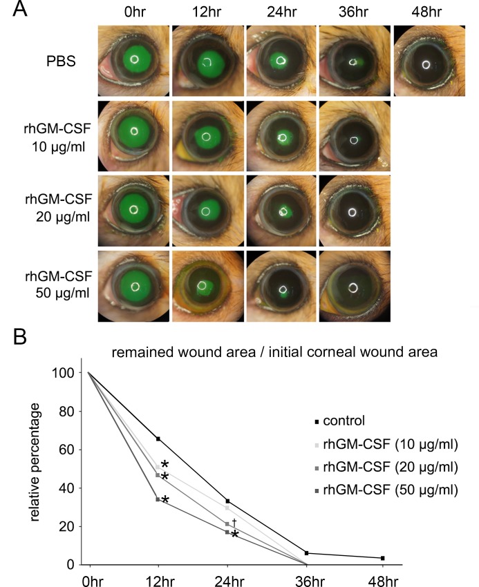
Fig 2. In vivo corneal epithelial wound healing.
(A) rhGM-CSF promoted accelerated corneal wound healing. Slit lamp photographs showed fluorescein staining of corneal epithelial wounds in the control and rhGM-CSF (10, 20, and 50 μg/ml) treatment groups at 0, 12, 24, 36, and 48 h after debridement. (B) Plotted data are the relative percentages of the corneal wound area at 12, 24, 36, and 48 h. The area of the initial corneal epithelial wound was normalized to 100%. The wound-healing rate of rhGM-CSF-treated eyes was much faster than that of untreated eyes. *P < 0.01 vs. control; †P = 0.02 vs. control.

