Fig. 6.

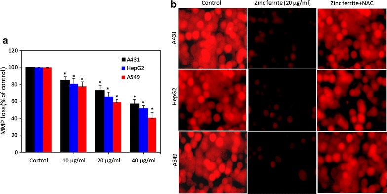
Zinc ferrite NPs induced MMP loss in A431, HepG2 and A549 cells. a Percentage change in MMP in all three cells after zinc ferrite NPs exposure at the concentrations of 0, 10, 20 and 40 µg/ml for 24 h. b Representative microphotographs showing MMP loss in A431, HepG2 and A549 cells after zinc ferrite NPs exposure at a concentration of 20 µg/ml for 24 h. Images were captured with a fluorescence microscope (OLYMPUS CKX 41). Data represented are mean ± SD of three identical experiments made in three replicate. *Statistically significant difference as compared to control (p < 0.05)
