
Fig. 3.
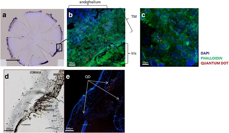
Tracking of MSCs after EVC in the AC. Whole flat-mounted cornea viewed with a digital fluorescence scanner (Nanozoomer) a. Confocal microscopy images at two different magnifications (b ×200, c ×800) of flat-mounted cornea showing cell nuclei (DAPI; blue), actin fibers (Phalloidin; green) and quantum dot (QD)-labeled MSCs (red) injected 21 days after EVC and found 23 days later at the time of sacrifice. Representative images of AC angle with contrast phase d and fluorescence microscopy e showing cell nuclei (DAPI; blue) and QD-labeled MSCs on the cornea endothelium, ciliary processes, and trabecular meshwork (TM) on a cryostat section (red). DAPI 4′,6′-diamidino-2-phénylindole