
Fig. 4.
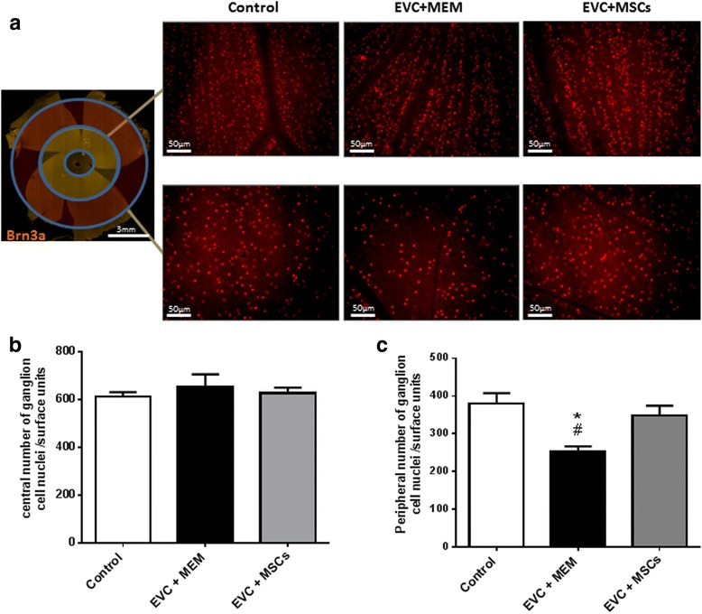
MSCs transplantation improves RGCs survival. Whole flat-mounted retina immunolabeled with brain-specific homeobox/POU domain protein 3A (Brn3a) antibody. Representative images (×200) of the peripheral or central area showing immunopositive staining of RGCs in noncauterized (Control) and cauterized eyes injected with MEM (EVC + MEM) or MSCs (EVC + MSCs) a Quantification of central b and peripheral c RGC densities per retina using automated nuclei counting in the control group (n = 5), EVC + MEM group (n = 5), and EVC + MSCs group (n = 5). The average RGCs density was obtained from eight peripheral and central images per retina and per area. Data expressed as mean ± standard error of the mean. *p <0.05 vs. control, #p <0.05 vs. EVC + MSCs. EVC episcleral vein cauterization, MEM minimum essential medium, MSCs mesenchymal stem cells