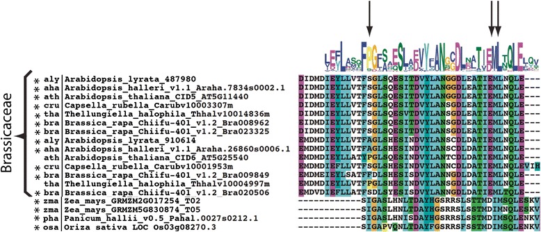
Fig. 6.

Alignment of the CUE motif of CID C proteins. The alignment region encompassing sequence LOGO #1 of Brassicaceae CID C2 sequences and of four CID C1 monocots is displayed; the alignment of all CID C sequences is shown in Additional file 8. The locations of important residues are highlighted by arrows (the invariant proline residue and a di-leucine motif). (*) indicates substitution of the invariant proline residue
