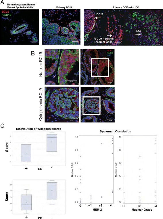
Fig. 10.

BCL9 may serve as a biomarker of high risk DCIS. a Immunofluorescence (IF) staining using anti-BCL9 antibody (red), anti-K5/K19 (green), and counterstained with 4′,6-diamidino-2-phenylindole (DAPI) (blue) in patient DCIS samples with and without invasion, and in normal adjacent human breast epithelial cells. b Representative IF images for patient DCIS samples with the above antibodies, demonstrating nuclear (top) and cytoplasmic (bottom) BCL9 expression patterns. c Statistical analysis of tissue sections from 28 patients purely with DCIS analyzed by IF using anti-BCL9 antibody. This analysis showed that the percentage of cells expressing nuclear BCL9 was significantly higher in DCIS that were estrogen receptor (ER)-negative (p = 0.004; Wilcoxon rank sum test), progesterone receptor (PR)-negative (p = 0.003; Wilcoxon rank-sum test), high human epidermal growth factor receptor 2(HER2) (Spearman correlation = 0.56; p = 0.002), and high nuclear grade (Spearman correlation = 0.49; p = 0.008). Median percentage (IQR) of cells positive for nuclear BCL9 for ER-positive samples = 8 % (1 %, 40 %); for PR-positive = 8 % (1 %, 35 %); ER-negative = 90 % (15 %, 95 %); PR-negative = 90 % (15 %, 95 %)
