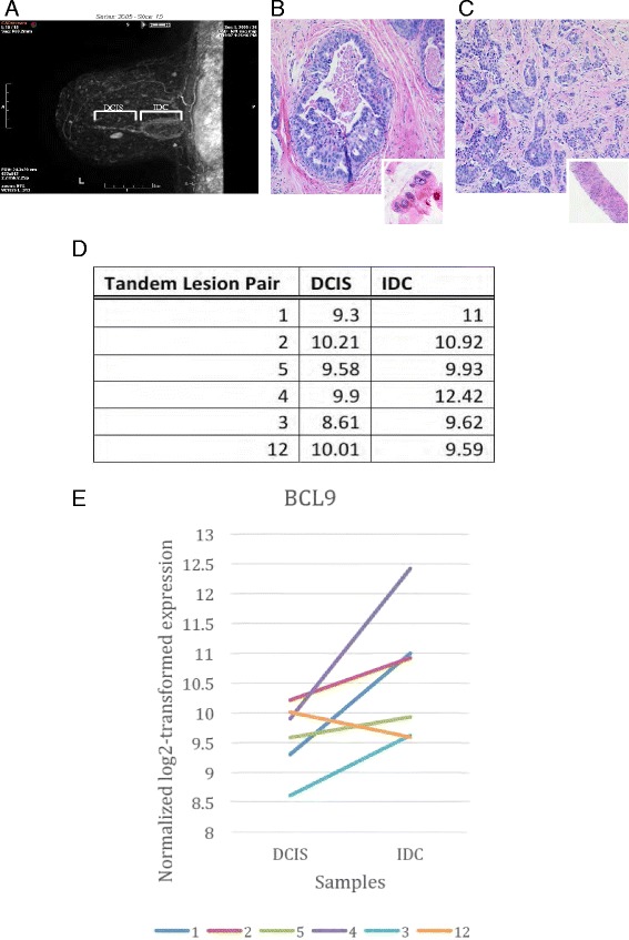
Fig. 2.

Analysis of RNA sequencing data of tandem lesions for BCL9 expression comparing DCIS to invasive ductal carcinoma. Radiographic (a) and H&E stain (b, c) images of a patient’s DCIS/IDC tandem lesion. a Three-dimensional ultrasound image of a tandem lesion. b H&E staining of a biopsy taken from the DCIS and (c) the IDC regions. Insets: lower magnification in b, c, respectively, demonstrating that the biopsies were highly pure and composed of either DCIS or IDC. d, e Normalized log2 transformed expression of BCL9 was plotted and tandem lesions between paired DCIS and IDC are connected by a line to indicate their relationship. The results indicate a significant increase in BCL9 expression in IDC samples compared to the DCIS lesions (p = 0.01)
