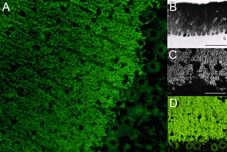
Fig. 3.

Binding of serum anti-mGluR1 from patients with ACA to rat (panels a and d) and mouse (panels b and c) cerebellum tissue sections. Patient antibodies were detected by use of goat anti-human IgG secondary antibodies labelled with Alexa Fluor®488 (panel a and d) or biotin (panel b: avidin/peroxidase; panel c: avidin/Texas red). While anti-mGluR1 strongly stain the molecular layer (a-d), staining of the PC somata varies in intensity depending on detection methods and antibody titres (compare panels a and c to panels b and d). Permission for panels b and c obtained from Massachusetts Medical Society. Copyright © (2000) Massachusetts Medical Society. Sillevis Smitt et al., Paraneoplastic cerebellar ataxia due to autoantibodies against a glutamate receptor. N Engl J Med. 2000; 342:21-27
