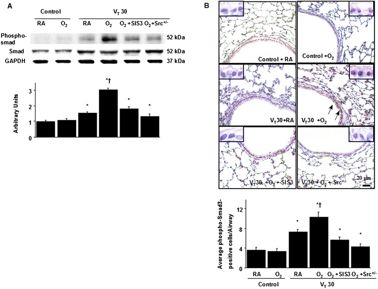
Fig. 4.

SIS3 and Src-deficient mice suppressed hyperoxia-augmented lung stretch-mediated Smad3 phosphorylation. a A Western blot was conducted by using an antibody that recognizes the phosphorylated Smad3 expression and an antibody that recognizes total Smad3 expression were from the lungs of nonventilated control mice and those subjected to a tidal volume of 30 mL/kg for 1 h with room air or hyperoxia. Arbitrary units were expressed as the ratio of phospho-Smad3 to Smad3 (n = 5 per group). b Representative micrographs (×400) with phosphorylated Smad3 staining of paraffin lung sections and quantification were from the lungs of nonventilated control mice and those subjected to a tidal volume of 30 mL/kg for 4 h with room air or hyperoxia (n = 5 per group). SIS3 2-mg/kg was given intraperitoneally 1 h before ventilation. A dark-brown diaminobenzidine signal identified by arrows indicates positive staining for phospho-Smad3 in the lung epithelium or interstitial, whereas shades of bluish tan signify nonreactive cells. *P < 0.05 versus the nonventilated control mice with room air; † P < 0.05 versus all other groups. Scale bars represent 20 μm
