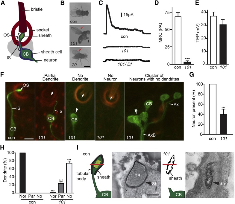
Figure 1.
mecA mutants have mechanosensory defects. (A) Illustration of the Drosophila mechanosensory organ. (B) mecA20 and mecA101 hold their wings erect (red arrow). Additionally, mecA101 cannot stand and cross their legs (black arrow). Bar, 2 mm. (C–E) mecA101 homozygotes and mecA101 hemizygotes have an abnormally low MRC (C and D) but normal TEP (E). Shown as mean ± SD; n ≥ 11. ***P < 0.0001. con, control; 101, mecA101. (F–H) mecA101 flies have abnormally short or missing sensory dendrites labeled by GFP-tubulin (F). Sensory outer segments labeled by GFP-tubulin are virtually absent in mecA101 (white arrow). Dendrites labeled by GFP-tubulin are often absent in mecA101 (arrowhead). Green: GFP-tubulin. Red: cuticle autofluorescence. Bar, 5 μm. (G) Quantification of the percentage of mechanosensory neurons in mecA101. Shown as mean ± SD; n = 250. ***P < 0.001. (H) Quantification of the percentage of abnormally short or missing dendrites in mecA101. Shown as mean ± SD; n = 250. ***P < 0.001. Nor: normal dendrite. Par: partially short dendrite. No: no dendrite. (I) Cross-sections of adult mechanosensory organ at the level of outer segment in control (con) and in mecA101 (101). The location of cross-sections is shown by the red bar. Bar, 0.5 μm. CB, cell body; TB, tubular body; Sh, dendritic sheath. (C–E and I) bw;st (control), bw;st;mecA101 (101); F, elav-Gal4;UAS-GFP-tubulin;MKRS/TM6B (control) and elav-Gal4;UAS-GFP-tubulin;mecA101 (101).

