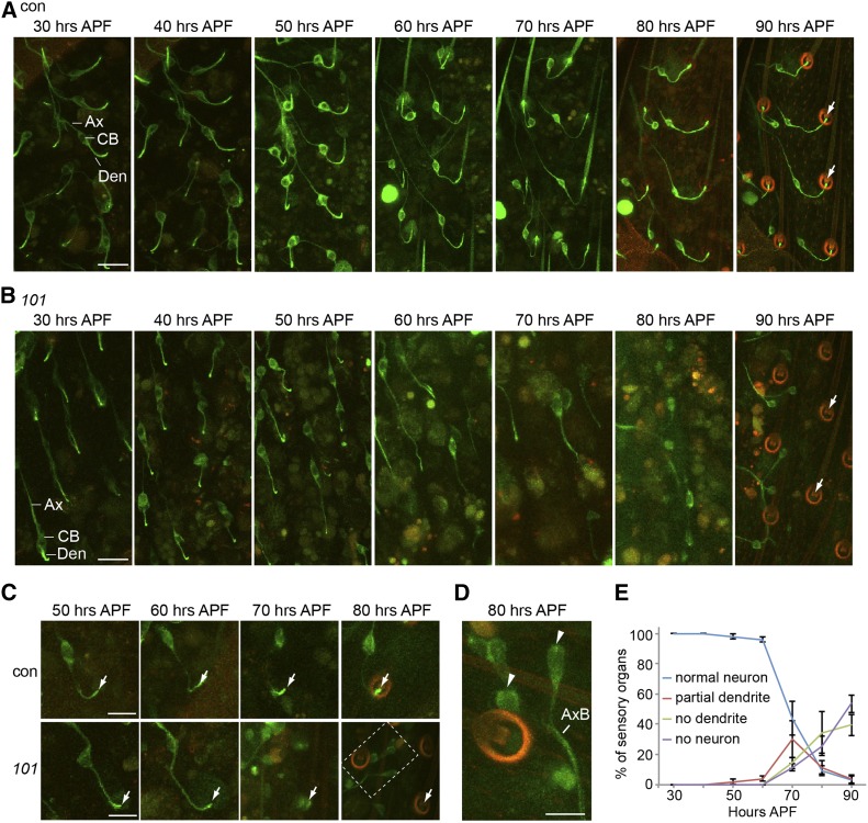
Figure 3.
Girdin is essential for sensory dendrite formation. (A and B) The development of sensory neuron in the thorax was monitored using confocal microscopy of GFP-tubulin. Starting from 30 hr APF, images were taken every 10 hr up to 90 hr APF in wild type (A) and girdin101 (B). Bar, 20 μm. (C) A single neuron was followed during development using live confocal microscopy of GFP-tubulin. Bar, 10 μm. A 488-nm laser with 5% power was applied for GFP fluorescent excitation; emission photons were collected from the wavelength 490–550 nm for the green and 620–750 nm for the red autofluoresce. The merge of this autofluorescence results in orange color. Ax, axon; CB, cell body; Den, dendrite. (D) Magnification of dashed-line box is at 80 hr APF in girdin101. Sensory outer segments labeled by GFP-tubulin are virtually absent in girdin101 (white arrow). Dendrites labeled by GFP-tubulin are absent in girdin101 (arrowhead). Green: GFP-tubulin. Red: cuticle autofluorescence. Bar, 5 μm. (E) Quantification of the percentage of normal neuron (having a dendrite with a cilium at the expected location for each stage), abnormally short or missing dendrites, or missing neurons in girdin101 over sensory organ development starting at 30 hr APF until 90 hr APF. Shown as mean ± SD; n = 250. (A–D) elav-Gal4;UAS-GFP-tubulin;MKRS/TM6B (control) and elav-Gal4;UAS-GFP-tubulin;girdin101 (101).

