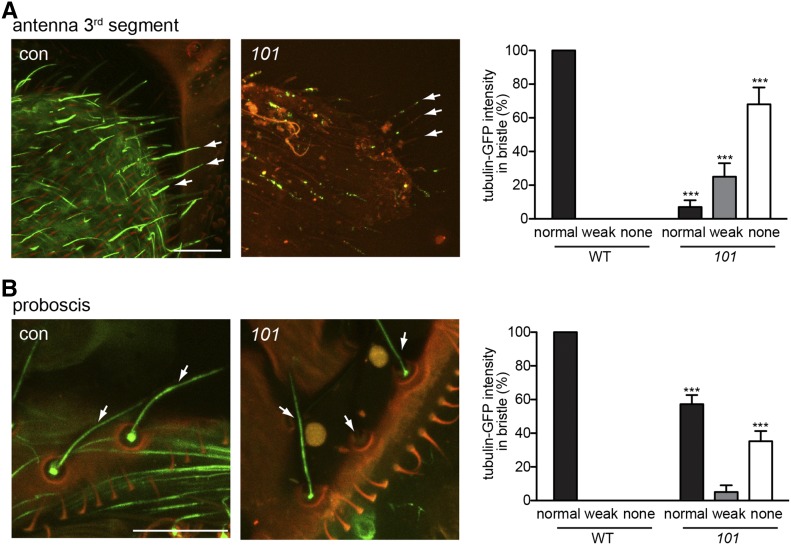
Figure 6.
girdin101 has abnormal ciliated dendrite in olfactory and taste organs. (A and B) Cilia intensity is abnormally weak or absent (white arrow) in olfactory neurons of the third segment of antenna (A) and taste neurons in proboscis (B) expressing GFP-tubulin. Quantification of GFP-tubulin intensity in bristles from the third segment of antenna shows that cilia are mostly absent or severely defected in girdin101 (A). Quantification of GFP-tubulin intensity in bristles from proboscis shows that 36% of cilia are absent in girdin101 (B). Bar, 20 μm. Shown as mean ± SD; n = 100. ***P < 0.0001. con, control; 101, girdin101. The green fluorescent emission photons were collected from the wavelength range of 490–550 nm with a laser power of 3% for the labeling of GFP-tubulin. Illumination of bristles causes the cuticle of the bristle shaft and socket to autofluoresce in red. The merge of this autofluorescence results in orange color. (A and B) elav-Gal4;UAS-GFP-tubulin;MKRS/TM6B (control) and elav-Gal4;UAS-GFP-tubulin;girdin101 (101).

