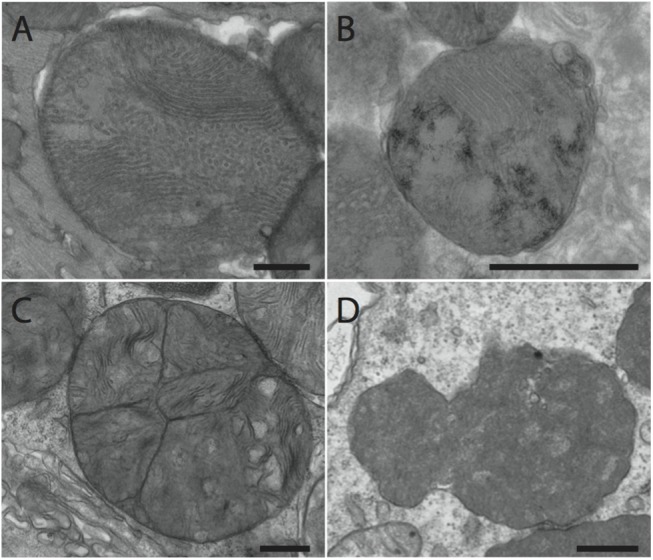
Fig 7. Representative images of abnormal mitochondria used to categorized TEM images from Abcc6 KO and WT CVB3-infected hearts.

Representative images were taken from heart sections from Abcc6 KO mice infected with 50pfu/g CVB3. (A) Disrupted cristae. (B) Fusion/fission events. (C) Hydroxyapatite deposition. (D) Abnormal mitochondria characterized by dense staining and irregular shape. Bar: 500nm
