. 2015 Aug 25;4:e08828. doi: 10.7554/eLife.08828

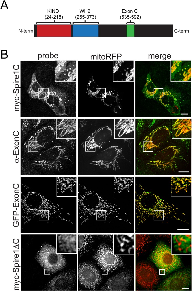
Figure 1. The Spire1 alternate exon ExonC is necessary and sufficient for localization to mitochondria.

(A) Full length Spire1C domain structure: The number ranges indicate the amino acid regions of the conserved domains probed in this study. (B) Spire1C localizes to mitochondria. myc-Spire1C: U2OS cells cotransfected with myc-Spire1C and mitoRFP show robust localization of myc-Spire1C to mitochondria. α-ExonC: U2OS cells stained with an antibody raised against ExonC (α-ExonC) and expressing mitoRFP show endogenous Spire1C labeling on mitochondria. GFP-ExonC: U2OS cells cotransfected with GFP-ExonC and mitoRFP show robust targeting of GFP-ExonC to mitochondria. myc-Spire1ΔC: U2OS cells cotransfected with myc-Spire1ΔC and mitoRFP show no specific targeting of myc-Spire1ΔC to mitochondria. All cells were fixed and primary antibodies were counterstained with Alexa-488 secondary antibody before imaging with confocal fluorescence microscopy. Scale bars: 10 μm. Inserts are magnifications of the boxed regions.

DOI: http://dx.doi.org/10.7554/eLife.08828.003

Figure 1.

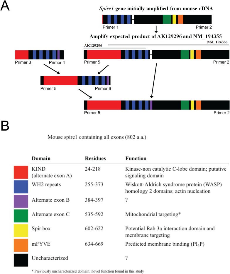
Figure 1—figure supplement 1. Construction of the complete Spire1C protein sequence as explained in detail in the ‘Materials and methods’.

Figure 1—figure supplement 1.

Spire1C gene amplified from mouse brain cDNA. (A) Initial construct based on NM_194355 was later combined with the sequence based on AK129296 to form full-length Spire1C protein of 802 amino acids with all known exons. (B) Spire1C diagram showing both characterized (red, blue, yellow, and orange) and uncharacterized (purple, green, and black) domains (indicated in the legend on the bottom).
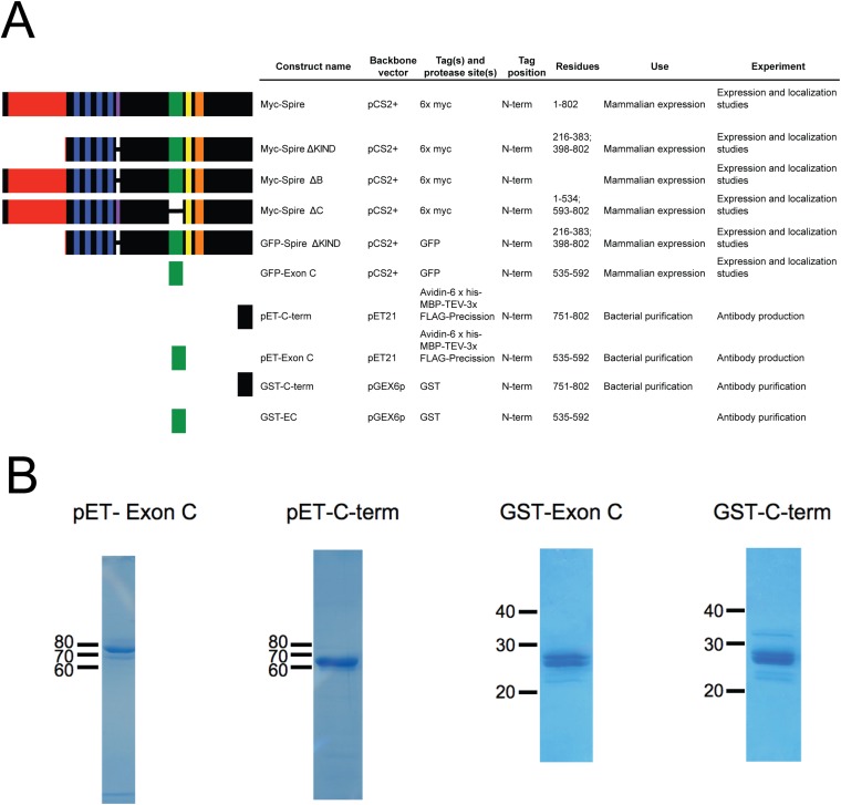
Figure 1—figure supplement 2. Constructs used to probe Spire1 function.

Figure 1—figure supplement 2.

(A) Spire1C constructs used in this study. Residue numbers are for the 802 a.a. full-length mouse Spire1C protein. (B) Coomassie-stained gels of purified proteins used for antibody production (pET constructs) and affinity purification (GST constructs).
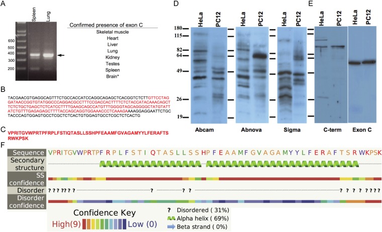
Figure 1—figure supplement 3. Spire1C contains a previously uncharacterized alternate exon of 58 amino acids.

Figure 1—figure supplement 3.

(A) Spire1 ExonC was found to be present in the tissues listed. *PCR from cDNA did not yield conclusive results, but the ExonC-containing gene was originally amplified from mouse brain cDNA. (B) Sequencing results from TOPO pCR2.1 vector using the M13 reverse primer. ExonC sequence is shown in red. (C) Translated protein sequence from red portion in C. (D) Representative immunoblots comparing commercial antibodies to Spire1 used to detect endogenous Spire1 in HeLa and PC12 cell lysates, 25 μg per lane. (E) Affinity purified Spire1 C-terminal antibody and ExonC antisera (both generated in this study) tested on HeLa and PC12 cell lysates, as in (A). Strikingly different protein bands are observed for the same cell type with different antibodies and between cell types with the same antibody. Molecular weight protein markers are the same size for each blot, though the positions vary slightly (as indicated). (F) The Phyre server was used to predict any secondary structural elements in ExonC. Phyre compiles structure predictions from three different algorithms (psipred, jnet, and sspro) to form a consensus prediction and probability for each structural element. Probability ranges from 1–10, with 10 being the most probable. Yellow amino acids are small/polar, green are hydrophobic, red are charged, and purple are aromatic + cysteine.