Fig. 3.

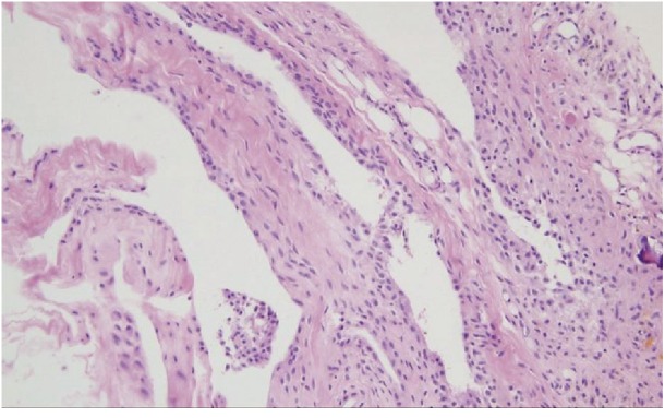
Histological appearance of the hemorrhagic synovial cyst showing synovial cell lining, fibroconnective tissue, neoangiogenesis, and hemosiderin microdeposits (H&E stain, ×200).

Histological appearance of the hemorrhagic synovial cyst showing synovial cell lining, fibroconnective tissue, neoangiogenesis, and hemosiderin microdeposits (H&E stain, ×200).