Fig. 1.

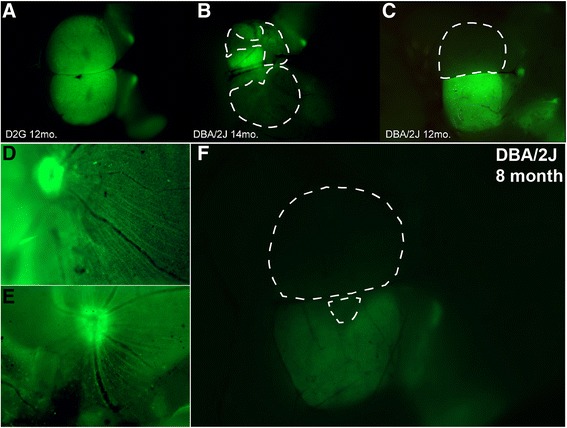
Coverage of cholera toxin-B (CTB) transport in the superior colliculus (SC). a–c Whole mount brain (cortex removed) of three mice demonstrating varying degrees of anterograde transport to major retinal targets that include lateral geniculate nucleus (LGN), pretectum (PT), and SC. a Colliculi taken from a 12-month-old D2G control mouse show intact axonal transport with full CTB coverage in both right and left SC. b Colliculi taken from a 14-month-old DBA/2J mouse show compromised transport; left SC has 56 % CTB coverage, right SC has 18 % CTB coverage. c Colliculi taken from 12-month-old DBA/2J mouse demonstrate both complete CTB dropout (left SC, 0 % CTB) and complete CTB coverage (right SC, 100 % CTB). Dotted lines indicate delineation used for microdissections of CTB-negative (i.e., transport absent) areas. d, e Whole mount retinas with successful CTB uptake via intravitreal injection. f Whole mount SC with areas of transport loss, corresponding to retinas in d and e. Retinal images (d, e) provide evidence that lack of CTB in SC is not indicative of failed intravitreal injection or lack of CTB uptake
