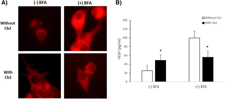
Fig. 3.

Intracellular VEGF levels in LGCs cultured with the dopamine receptor 2 agonist, Cabergoline. a Representative immunofluorescence images of intracellular VEGF staining in fixed and permeabilized LGCs (n = 6) pre-incubated with BFA and then incubated in the presence or absence of Cabergoline (Cb2) with hCG plus or minus BFA. b VEGF immunofluorescence quantification as assessed by Matlab, expressed as eu/pixel. In the absence of BFA (−), intracellular VEGF levels in Cb2-treated LGCs were higher than the VEGF levels in Cb2-untreated LGCs. However, in the presence of BFA (+) the intracellular VEGF levels in Cb2-treated cells were lower than vehicle-treated cells. *p < 0.05 compared to Cb2-untreated conditions
