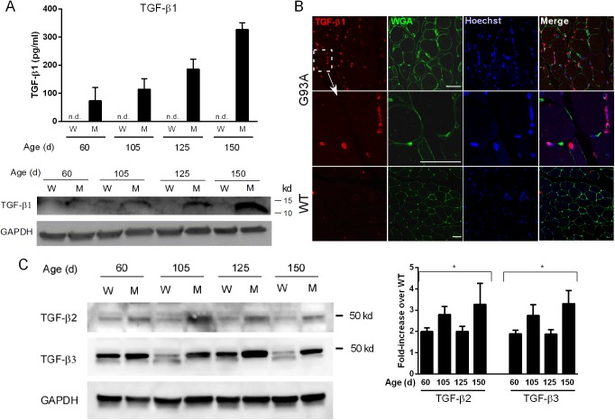
Fig 4. TGF-β protein is elevated in G93A mouse muscle.
(A) Muscle lysates from G93A SOD1 mice (M) and non-transgenic littermates (W) were assessed for TGF-β1 by ELISA and Western blot. ELISA values were determined by comparison to a standard curve. A representative Western blot of the lysates (under reducing conditions) shows expression of mature TGF-β1. ELISA data represent the mean ± SE of 3 mice. The Western blot was repeated once with the similar results. (B) Confocal photomicrographs of G93A or WT muscle sections using a TGF-β1 antibody. WGA, wheat germ agglutinin. Size marker = 50 microns. (C) A representative Western blot of TGF-β2 and β3 in mouse muscle showing increased levels of the unprocessed peptide in mutant versus control samples. Quantitative densitometry of TGF-β ligands was performed on three Western blots from three independent mouse samples. Data are shown as fold-increase over WT controls. *p < 0.05 for each age.

