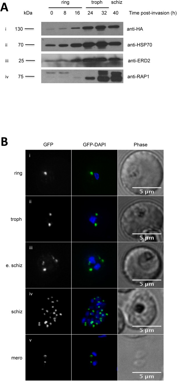
Fig 2. PfPRP2-RFA is constitutively expressed throughout the erythrocytic cycle.
(A) Western blots on parasite extracts from tightly synchronous parasites show that PfPRP2-RFA is detected throughout the blood stages (Ai, anti-HA) similar to the constitutive cytosolic protein HSP70 (Aii) and the constitutive Golgi protein ERD2(Aiii). The rhoptry protein RAP1 is detected in very early and late stage parasites (Aiv). (B) Dynamics of the localization of PfPRP2-RFA throughout the asexual stages. In ring stage parasites, a single dot of fluorescence is seen close to the nucleus (Bi). In trophozoites, the dot starts to multiply, prior to nuclear division (Bii). Multiplication of the PfPRP2-RFA signal continues once nuclear division is undertaken (Biii, Biv). In free merozoites, each parasite inherits one locus of PfPRP2-RFA fluorescence (Bv). The fluorescence of PfPRP2-RFA is pseudocolored in green and the nuclei of parasites were stained with DAPI (blue). Scale bar represents 5μm. Troph: trophozoite; E. schiz: early schizont; Schiz: schizont; Mero: merozoite.

