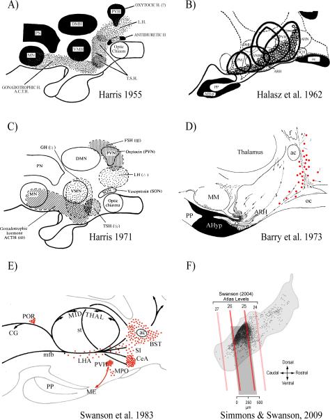
Figure 1.
Six maps from 1955 to 2009 show the hypothalamic locations of regions containing neuroendocrine neurons and pituitary hormone control mechanisms. They illustrate the dramatic improvement in the resolution of these representations since 1955.
A) Diagram of a midline sagittal section through the hypothalamus and pituitary gland. The stippled areas indicate the sites where electrical stimulation or lesions have resulted in changes of pituitary secretion. Abbreviations: ACTH, adrenocorticotrophic hormone; DMH, dorsomedial nucleus of the hypothalamus; LH, luteinizing hormone; MN, mammillary nuclei; PN, ‘posterior nucleus’; PVH, paraventricular nucleus of the hypothalamus; TSH, thyroid stimulating hormone; VMH, ventromedial nucleus of the hypothalamus. Adapted from Harris [1955b].
B) The hypophysiotropic area of the hypothalamus. The solid black line represents the borders of five relatively midline pituitary grafts in which considerable cellular integrity was maintained despite their ectopic site. The dotted circles are the locations of periodic acid-Schiff-positive basophils. Abbreviations as A) except: AHN, anterior hypothalamic area; AHyp, adenohypophysis; ARH, arcuate nucleus; oc, optic chiasm; PM, premammillary nucleus; PP, posterior pituitary; SCH, suprachiasmatic nucleus. Adapted from from Halasz et al. [1962].
C) As A), except both stippling and cross-hatching are used to indicate the sites where electrical stimulation or lesions have resulted in changes of pituitary secretion. Adapted from Harris [1972]
D) General diagram of the system of LRF (GnRH)-producing cells in a paramedian sagittal section of guinea-pig hypothalamus. Red dots show specifically immunoreactive perikarya. The dotted lines show the pathway of LRF axons (the arrows showing direction of transport). Abbreviations as A) and B), except: ac, anterior commissure; MM, median mammillary nucleus. Adapted from Barry et al. [1973].
E) A schematic illustration of the major CRF (CRH)-stained cell groups (red dots) and fiber systems (black lines) represented on a sagittal view of the rat diencephalon. Abbreviations as A) B), and D) except: BST, bed nucleus of the stria terminalis; CeA, central nucleus of the amygdala; CG, central gray; LHA, lateral hypothalamic area; ME, median eminence; mfb, medial forebrain bundle. MPO, medial preoptic area; POR, periocularmotor nucleus; SI, substantia innominata; st, stria terminalis. Adapted from Swanson et al. [1983].
F) The location of individual CRH neuroendocrine neurons (black dots) shown on a sagittal view of the rat paraventricular nucleus of the hypothalamus (PVH; light gray outline). The oblique red/dark gray lines show the corresponding positions of four atlas levels (24–26) from Swanson [2003]. Note that the vast majority of CRH neurons are found in the dorsal aspect of the PVH between levels 25 and 26 (darker gray box). In the coronal plane, most CRH neuroendocrine neurons are found in the dorsal zone of the medial parvicellular (mpd) part of the PVH. Adapted from Watts & Khan et al. [2013] using results originally published in Simmons & Swanson [2009].

