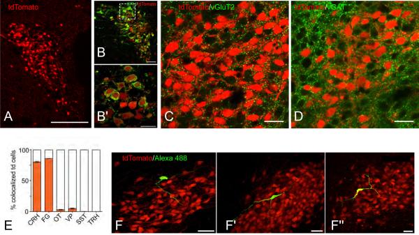
Figure 3.
Anatomical distribution and CRH protein expression in tdTomato labelled cells in the paraventricular nucleus of the hypothalamus (PVH) of Crh-IRES-Cre;Ai14 mice.
A) Confocal image (20× magnification) of the PVH in a Crh-IRES-Cre;Ai14 (tdTomato, red) mouse (level 61 of Dong [2007]). B) Confocal image (40×) of a colchicine-treated Crh-IRES-Cre;Ai14 mouse PVN. Immunostaining for corticotropin-releasing hormone (CRH) is shown in green. B’) Higher magnification (100×) of the box inset in (B). Confocal images of the PVH in Crh-IRES-Cre;Ai14 (tdTomato, red) mouse labeled for vGluT2 (C, green), or vGAT (D, green). Note the numerous close appositions (yellow) between tdTomato (CRH) neurons and glutamaterigic (vGlutT2) and GABAergic (vGAT) elements. E) Bar graph showing the percent of tdTomato positive cells that coexpress various neuropeptides, each from n=5 colchicine-treated mice. Abbreviations: CRH, corticotropin-releasing hormone; FG, fluorogold; OT, oxytocin; SST, somatostatin; TRH, thyrotropin-releasing hormone. F) Confocal Images (60× magnification), from 3 mouse PVH sections (F – F”), showing morphology of single tdTomato neurons (red) filled with Alexa488- (green) and biocytin (yellow) during whole-cell patch clamp recordings.
Scale bars are 100 μm (A), 50 μm (B, F - F”), and 20 μm (B’, C, and D). Panels A), B), E), and F) are adapted from Wamsteeker et al. [2013]. Panels C) are D) are unpublished photomicrographs from the author's laboratory (C.S. Johnson & A.G. Watts).

