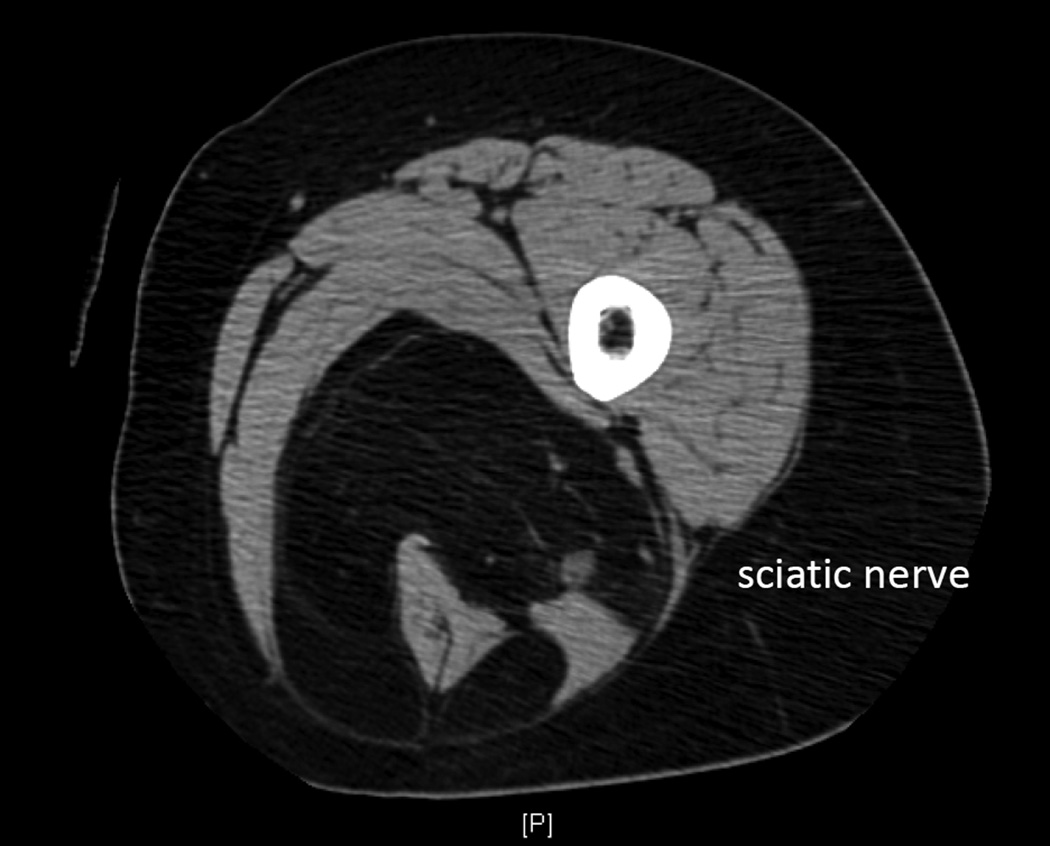
Figure 3.
Cross sectional imaging (CT) demonstrating a well-differentiated liposarcoma in the posterior compartment of the thigh, encasing the sciatic nerve and hamstring muscles. On both CT and MRI, well-differentiated liposarcoma has imaging characteristics almost identical to normal fat. Enhancing septations may be present and nodularity appears in the context of dedifferentiation.

