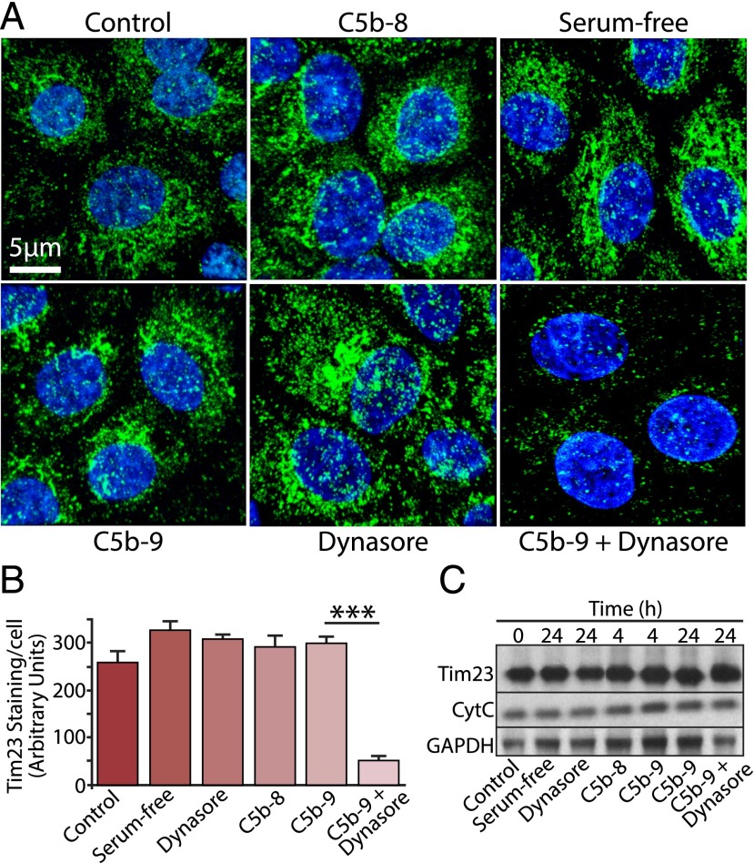
FIGURE 4.
Persistent exposure to C5b-9 leads to mitochondrial perturbation. (A) RPE cells were cultured on Transwells for 24 h and exposed to a variety of experimental conditions, as indicated in the figure. Cells were fixed and immunostained for the mitochondrial marker Tim23 (green) and DAPI (blue). (B) Data are expressed as mean ± SEM (n = 4 images/experiment from three independent experiments). Quantitative analysis of Tim23 staining revealed a significant reduction (***p < 0.001) in staining intensity in cells treated with C5b-9 and Dynasore versus C5b-9 or Dynasore alone. (C) Whole-cell lysates were prepared from RPE cells cultured under the same set of conditions as above for the times indicated and Western blotted for Tim23, cytochrome C (CytC), and GAPDH as a control. No discernable difference was noted in the band intensities for either mitochondrial protein, under any of the different conditions.

