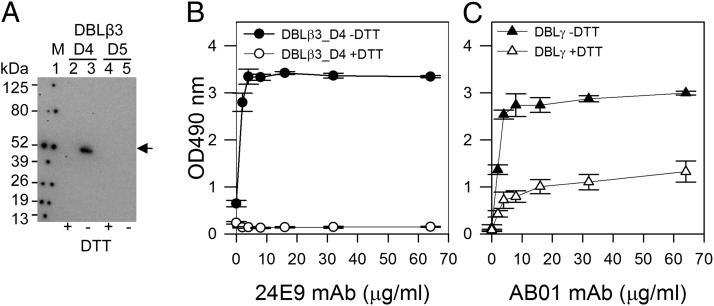
FIGURE 5.
24E9 mAb recognizes a conformation epitope on PFD1235w DBLβ3_D4. (A) Western blotting of PFD1235w DBLβ3_D4 (D4) and DBLβ3_D5 (D5). +DTT (reduced), −DTT (nonreduced). Lane 1, Prosieve protein marker (M) visualized by phosphorescent paint as dots. Lanes 2 and 3, DBLβ_D4 (±DTT). Lanes 4 and 5, DBLβ3_D5 (±DTT). Arrow shows nonreduced DBLβ3_D4 (lane 3) recognized by 24E9 mAb. (B) 24E9 mAb ELISA reactivity against reduced (+DTT) and nonreduced (−DTT) PFD1235w DBLβ3_D4. (C) AB01 mAb ELISA reactivity against reduced (+DTT) and nonreduced (−DTT) DBLγ of PFD1235w. Mean OD values are shown for three independent experiments. Error bars indicate SD.

