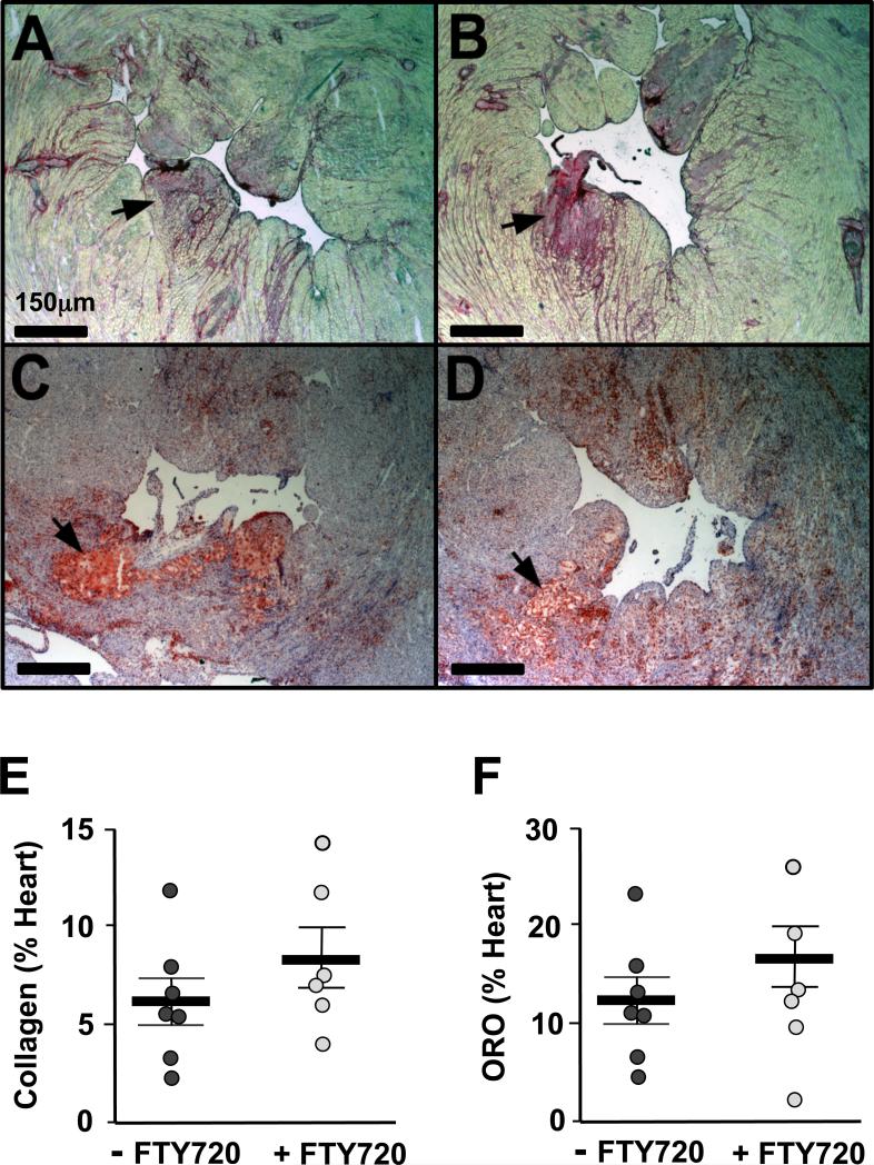
Figure 6.
Images of heart sections showing the left ventricular chamber stained with Sirius Red (A and B) and Oil red O (C and D) of ApoeR61h/h/SRB1–/– mice fed the HFD for 3.5 weeks with FTY720-treatment or not (n=7-8 mice/group). Quantification of collagen (E) and neutral lipid (F) deposited in the myocardium. Black arrows identify the area with clusters of collagen or neutral lipid. Data are mean ± SEM. ORO = oil red O

