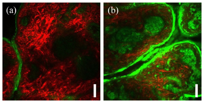
Fig. 4.

Representative microscopy images of porcine skin incubated for 17 days in a) PBS control solution and b) 0.165 M ribose solution. Shown are composite images of multiphoton autofluorescence (green) and second harmonic generation (red) channels. Images were acquired at the depth of 35 µm. Scale bar is 50 µm.
