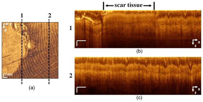
Fig. 10.

(a) En face OCT image in which dashed lines 1 and 2 correspond to the cross-sectional images in (b) and (c), respectively. The cross section line 1 in (a) includes scar tissue and a portion of healthy tissue. On the cross-sectional image in (b), the thickness of the epidermis in the scar portion is significantly reduced and the morphology of the skin layers is altered. (c) The cross-sectional image of healthy tissue. The dermis−epidermis junction, sweat ducts, and the overall thickness of the epidermis, which is constant along the x axis, can be seen. x-axis scale bar: 1 mm; z-axis scale bar: 100 µm.
