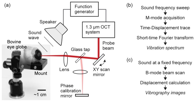
Fig. 2.

(a) Schematic of the experimental setup. The sound wave emitted from a loudspeaker is directed toward the eyeball, and the motion of the cornea is captured and recorded by the OCT system. A needle syringe connected to a water column (not shown in this picture) is placed into the globe, by which the IOP was set to 6 mmHg. (b) Signal processing sequence for the measurement of vibration spectrum at a fixed spatial location in the cornea. (c) Signal processing sequence to generate vibrography images at a specific sound frequency.
