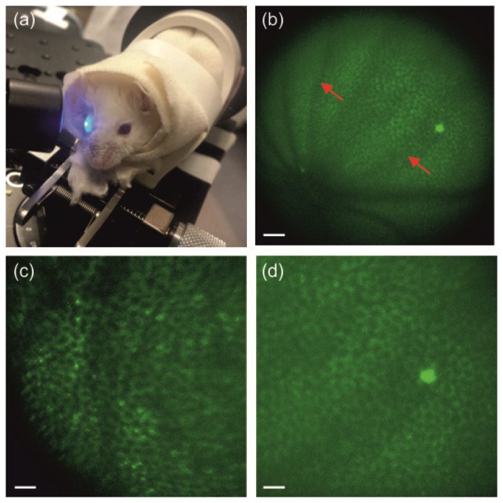
Fig. 3.

Two-photon imaging of the RPE in living WT and Rpe65–/– mice. (a) Mouse eye illuminated with the newly designed periscope. (b) Image of the RPE in the WT albino mouse. (c), (d). Enlarged images of the RPE in Rpe65–/– (c) and WT (d) albino mice. Excitation wavelength: 730 nm. Mean power at the cornea: 20 mW in (c) and 25 mW in (b) and (d). Scale bars: 100 µm in (b), and 50 µm in (c) and (d). Red arrows indicate shadows from retinal vasculature.
