Fig. 1.

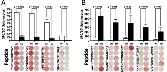
ERAP1 reshapes the immunodominant focus by the creation and destruction of epitopes. C56BL/6 mice (n = 5) or ERAP1−/− mice (n = 5) were immunized I.M. in the tibialis anterior with Ad5-Clostridium difficile-TA as described in Methods. At day 14, mice were sacrificed and splenocytes were harvested and stimulated with 2 μg per well of the listed 15-mer peptides. Splenocytes were then analyzed for IFN-γ production using IFN-γ ELISpot. From the 84 peptide library, the peptides that generated a strong response (100 or more spots) and showed an average of at least 100 less spots in the other strain of mouse were selected and repeated. (A) Peptides that had a very high response in WT mice compared with ERAP1−/− mice. (B) Peptides that had a very high response in ERAP1−/− mice compared with WT mice. Bars represent the mean number of spot-forming cells per 106 splenocytes ± SEM. Below are representative wells. All wells are shown in the order they appeared on the plate and all paired sets of wells are from the same plate. A P value < 0.05 was statistically significant. The figure is representative of two separate experiments.
