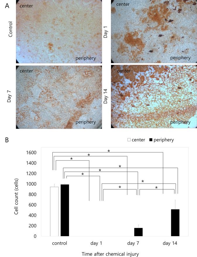
Fig 4. Vital staining of rabbit corneal endothelium with alizarin S red and trypan blue after injection of 0.1 mL of 0.05 N NaOH.
A. Corneal endothelial cells (CECs) in both the central and peripheral areas show damage at day 1. At day 7, CECs appeared at the periphery and scanty cells existed at the midperiphery and center 7 days after injury. The peripheral cells began to migrate from the far periphery and to recover from damage. At day 14, the dense small cells were observed at the periphery, the loose large irregular cells appeared at the midperiphery and no cells were still observed at the center of the cornea. The cell integrity of the peripheral cells recovers while cells migrate from the periphery to the center and also recover. B. Cell counts at ×400 magnification. Corneal endothelial cell numbers in the center and in the periphery are significantly lower compared to the control during the entire observation period (p < 0.001 in the center at all time points; p < 0.001 at day 1, p < 0.001 at day 7 and p = 0.012 at day 14 in the periphery). No corneal endothelial cells are present in the periphery or in the center 1 day after injury. Corneal endothelial cells in the periphery increase significantly over time (p < 0.001, one-way ANOVA). Corneal endothelial cells are detected in the periphery 7 days after injury and increase until day 14 (p = 0.022 compared to day 7), although they are lower compared to the number on day 1 (p = 0.014 and p = 0.010). Corneal endothelial cell counts in the center are not observed until day 14. * Statistically significant by Student’s t-test

