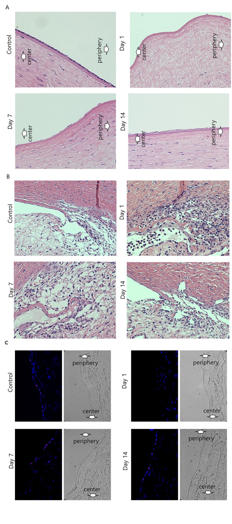
Fig 5. Microscopic images of hematoxylin and eosin staining in corneal endothelium (A) and trabecular meshwork (B), and immunofluorescent staining for the proliferation marker Ki-67 (C).

A. With hematoxylin and eosin staining, no cells were observed on the center and periphery of Descemet's membrane 1 day after injury. At day 7, corneal endothelial cells (CECs) were detected at the periphery, but not at the center. At day 14, CECs were detected at the mid-periphery and the periphery. B. With hematoxylin and eosin staining, a lot of inflammatory cells were infiltrated around trabecular meshwork 1 day after injury. At day 7, inflammatory cells disappeared and at day 14, trabecular meshworks were recovered much. C. Ki-67 staining was performed to assess the proliferative cells. Corneal sections were immunostained for Ki-67 (Red) and counterstained with 4',6-diamidino-2-phenylindole (DAPI; blue). Phase contrast images reveals the actual sections. No cells were observed on the center and periphery of Descemet's membrane 1 day after injury. At 7 and 14 days after injury, the cells expressing Ki-67 were observed at the periphery of the cornea. Magnification X 400.
