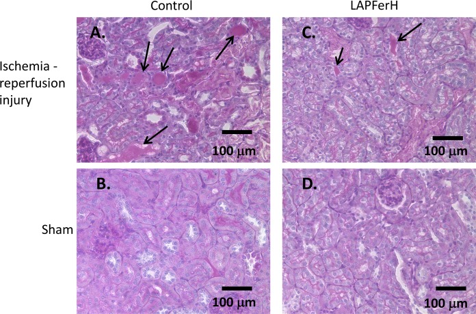
Fig 2. Histopathologic examination of kidney sections reveals changes consistent with ischemia-reperfusion injury.
Mice were subjected to 45 min renal ischemia to the right kidney and then allowed to recover for 24 hr. The contralateral left kidney was sham treated. Periodic acid-Schiff (PAS) stained kidney sections are shown in panels A-D]. Note the degree of loss of brush border and tubular sloughing in the cortical kidney (arrows) following ischemia-reperfusion injury is greater in the Control (A.) compared to the LAPFerH (C.) mouse. Normal tubular morphology is observed in the sham kidneys of both Control (B.) and LAPFerH animals (D.)

