
Figure 2.
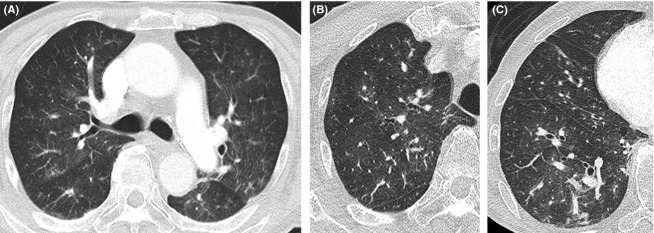
Chest CT (computed tomography) showed ground-glass opacities in both lung fields (A). High-resolution CT showed small nodules and ground-glass opacities in the upper lung field (B) and small nodules, ground-glass opacities, and dilation of the pulmonary arteries in the lower lung (C).