
Figure 4.
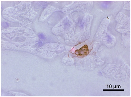
Light microscopic picture of a slice of guinea pig heart perfused with a bolus of 3 × 10⁶ human polymorphonuclear neutrophilic granulocytes, prestimulated with formyl-Meth-Leu-Phe. The heart was fixation-perfused with formalin, sliced and stained with antibody against human elastase. The brown colouration identifies elastase in granules within a PMN attached to the wall of a small venule and beginning to spread out along the endothelial surface. For details, see [26,134]