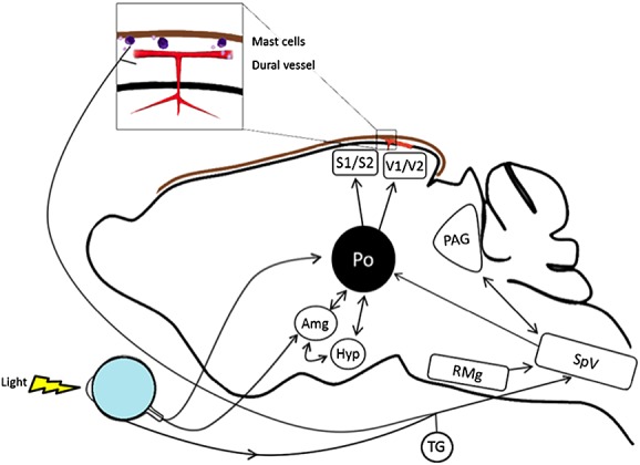
Figure 2.

Potential sites of CGRP action in light-aversive behaviour. The rodent brain is shown schematically, with arrows indicating pathways between relevant nuclei and input from the trigeminovascular system and light detected by the eye. These nuclei have calcitonin gene-related peptide (CGRP) receptors and respond to CGRP to modulate either affective behaviour or spinal trigeminal nucleus activity. Not all pathways are shown in this simplified presentation. Abbreviations are as follows: Amg, amygdala; Hyp, hypothalamus (which refers to the A11 nucleus and paraventricular nucleus); TG, trigeminal ganglion (which includes neurones and satellite glia); PAG, periaqueductal gray; Po, posterior thalamic nuclei (which include the posterior, ventroposteromedial and lateral posterior thalamus); Rmg, raphe magnus nucleus; SpV, spinal trigeminal nucleus; S1, S2, somatosensory cortex; V1, V2, visual cortex. Dural mast cells and blood vessels with associated trigeminal fibres and Schwann cells (not shown) are also indicated (brown line represents the dura)
