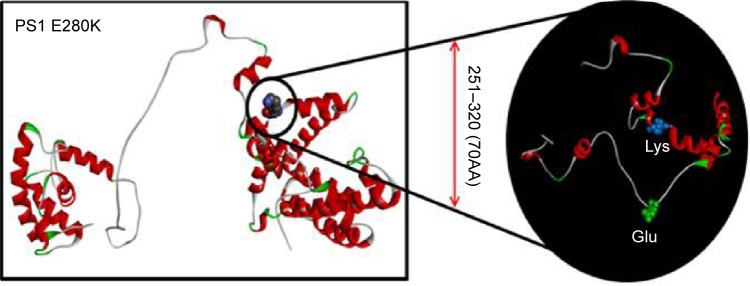
Figure 3.

In silico 3-D modeling for PS1 E280K, comparing to the normal PSEN1.
Notes: In the black circle, the differences between the normal and mutant PSEN1 proteins were highlighted. Glutamic acid was labeled with green since lysine was labeled with blue. Our prediction suggested that the mutation might result in significant changes in PSENl by generating extra helices in the loop structure.
Abbreviations: PSEN1, presenilin 1; 3-D, three-dimensional.
