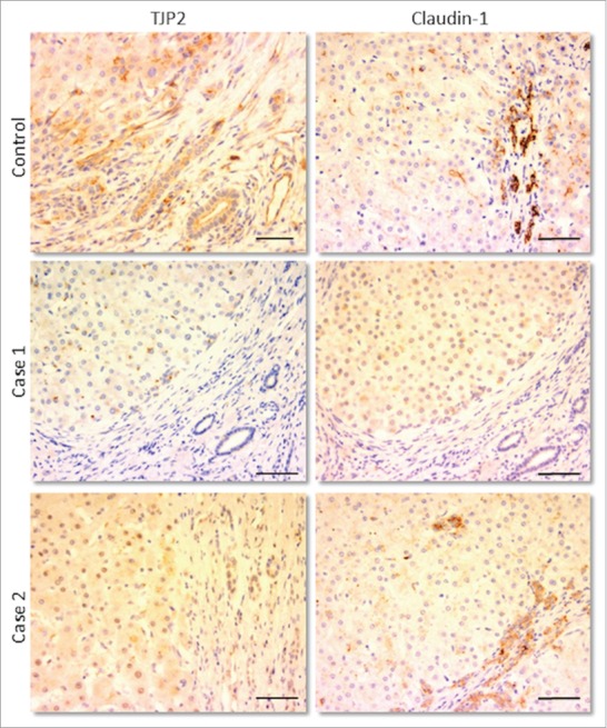
Figure 2.

Immunohistochemical staining for TJP2/ZO-2 (left column) and claudin-1 (right column) in livers of control sample and of 2 patients both having distinct protein-truncating mutations in TJP2. In each image, 2 areas can be distinguished: the hepatic parenchyma (top/left) and the portal tract (bottom/right), where bile ducts are visible. In the patients, no expression of TJP2 is present in either site, while that of claudin-1 appears reduced, more so in the parenchyma. Scale bars: 100 μm.
