
Figure 4.
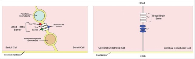
A simplified diagram illustrating the morphological differences between the blood‐testis barrier (BTB) and the blood‐brain barrier (BBB). (A) In the BTB, tight junctions (TJs) coexist with basal ectoplasmic specializations (ES), basal tubulobulbar complexes (TBC), and desmosome‐like junctions. (B) In the BBB TJs are restricted to the apical surface of the endothelium, sealing the intercellular space, with adherens junctions (AJ) located immediately below. Modified from ref. 30.