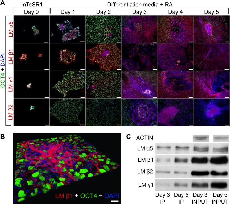
Fig 2. The expression of laminin α5, β1, β2 and γ1 chains in differentiating RA-treated hESC.
(A) Immunofluorescence analysis of laminin (LM) chains α5, β1, β2 and γ1 and OCT4 in RA-treated hESC. Laminin chains (red) and OCT4 (green) were detected with appropriate antibodies. Cell nuclei were labeled with DAPI (blue). Scale bar: 100 μm. (B) Multilayer confocal microscopy was used to visualize the LM β1 chain (red) localization and OCT4 (green) expression in RA-treated hESC. Cell nuclei were labeled with DAPI (blue). Scale bar: 20 μm. (C) Immunoprecipitation of laminin-511 and -521 from RA-treated hESC. The protein complexes were immunoprecipitated using laminin α5 chain-specific antibody. The laminin α5, β1, β2 and γ1 chains were detected by Western blot analysis using corresponding antibodies.

