. 2012 May 31;16(2):70–74. doi: 10.14701/kjhbps.2012.16.2.70

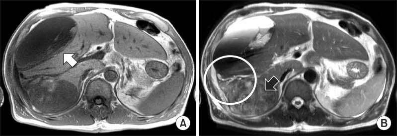
Fig. 2. Magnetic resonance imaging study shows (A) the large tumor of posterior lobe in the liver show heterogeneous lower signal intensity in T1 weighted image (white arrow) and (B) heterogeneous high signal intensity with necrosis and hemorrhage on mass in T2 weighted image (black arrow). It was associated with focal dilation around the bile duct and hepatic capsular retraction of the posterior section in the liver. The large hematoma of the right anterior section was connected with the heterogenous tumor of the right posterior section by high signal intensity in the right perihepatic space that was suspected as hemorrhage (white circle).

Fig. 2