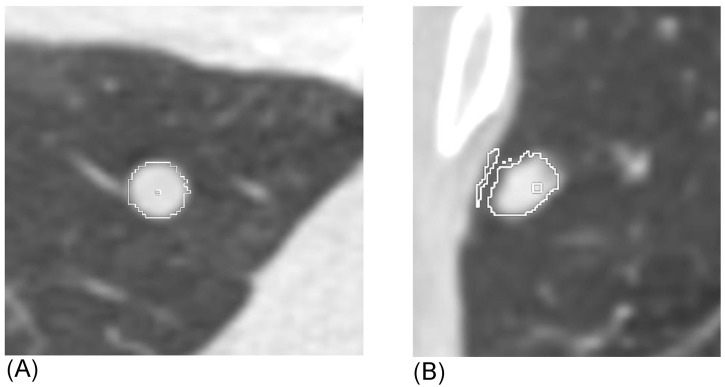
Fig 1. Example of automated volume extraction of a calcified nodule by VCAR software.

(A) Successful extraction: Note the contour fit to the nodule perimeter in multiple projections appears to correlate with the visual nodule outline. (B) Unsuccessful extraction: Note the clear erroneous border that includes adjacent chest wall in this subpleural nodule.
