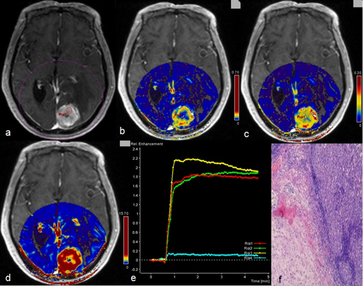
Fig 5. Description of metastatic tumor by multiple parameters from DCE-MR.
A 72 years old male had a metastatic tumor in the left occipital lobe from renal cell carcinoma. Contrast-enhanced T1-weighted MRI showed that the tumor is obviously enhanced with severe peritumoral edema (a) and the corresponding Ktrans (b), Ve (c) and iAUC (d) maps were also showed high value inside tumor parenchyma except central necrosis; concentration-time curve of the tumor (e), it manifested as plateau; brain metastases from renal cell carcinoma (f), (Hematoxylin- Eosin(HE) ×4).

