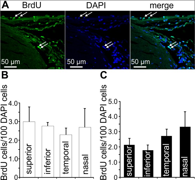
Figure 2.

BrdU-positive cells in the chamber angle. (A) Immunohistochemical staining for BrdU (green) in cells of the TM outflow pathway. Nuclei are stained with DAPI (blue). Arrows indicate BrdU-positive cells in Schlemm's canal endothelium and in the region of Schwalbe's line. (B, C) Quantification and statistical analysis of BrdU-positive cells in the different quadrants of group 1 ([B], chronic BrdU) and group 2 ([B], chronic BrdU and long-term retention) eyes. Means ± SEM are shown.
