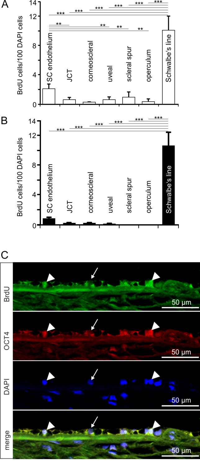
Figure 4.

BrdU-positive cells in Schwalbe's line region. (A, B) Relative number of BrdU-positive cells in Schwalbe's line region in comparison with that in the different regions of the TM outflow pathways in group 1 (A) and group 2 (B) eyes. Means ± SEM are shown, **P < 0.01. ***P < 0.001. Due to structural damage at the Schwalbe's line, one eye could not be included in this analysis. (C) Immunohistochemical staining of Schwalbe's line cells in a group 2 eye for BrdU (green) and OCT4 (red). Nuclei are stained with DAPI (blue). Arrowheads indicate BrdU/OCT4-positive cells in Schwalbe's line region, while the arrow points toward a BrdU/OCT4-negative nucleus that is stained with DAPI.
