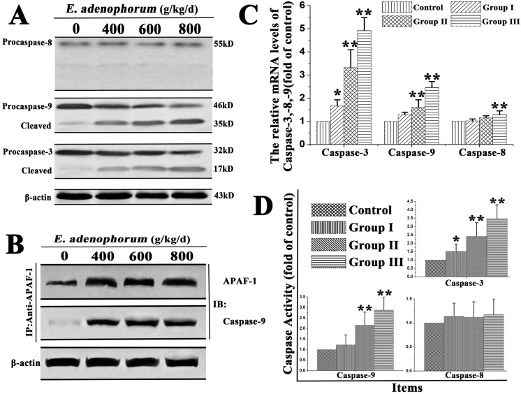
Fig 3. E. adenophorum-induced apoptosis is mediated by activation of caspase-9, caspase-3.
(A) The protein levels of procaspase-3, -8, -9 and the cleaved form of them. The expression of apoptosis-related proteins, including caspase-3, -9, -8 were shown with β-actin as a control, were detected by Western-blot analysis.(B) E. adenophorum induced apoptosome formation. Protein extractions from renal cells were collected and then used in immunoprecipitation assays against Apaf-1. The level of caspase-9 was detected by western blot to indicate the formation of apoptosome complex. (C) The relative mRNA levels of caspase-3, -8 and -9. The Saanen goat was treated with different dose of E. adenophorum for 3 months and the mRNA of renal cells was extraced and used for qRT-PCR assay. E. adenophorum induced the activiation of caspase-3, -9.(D) Caspase activities in renal cells. BCA assay was used to equal protein amounts and the enzymatic activities of caspases-8, -9, and -3 were measured using the colorimetric assay kits. Data are presented with the means±SD and mean values of three independent experiments. *p<0.05 and **p<0.01, compared with the control group.

