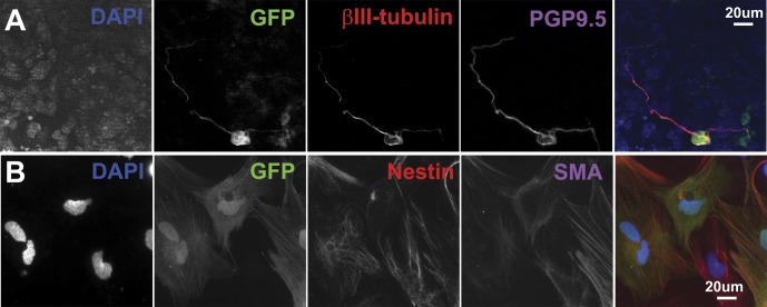
Fig 8. Multipotentiality of P/G-iPSC-derived GFP-positive cells.
Neural primed cells were induced to form spherical aggregates in the presence of CHIR99021. After 4 days of culture, the aggregates were cultured further to allow adhesion onto fibronectin-coated glass coverslips for 7 days in N2/B27 medium in the presence of CHIR99021. A, Immunostaining for GFP (green), βIII-tubulin (red), and PGP9.5 (purple). In B, the aggregates were dissociated into single cells, quickly reaggregated and cultured in N2 medium (DMEM/F12 plus N2 supplement) containing 20 ng/ml bFGF and 2 μg/ml heparin for 3 days to form neurospheres, and then cultured under differentiation conditions. Immunostaining for GFP (green), nestin (red), and α-smooth muscle actin (SMA, purple). Blue represents DAPI staining. Scale bars indicate 20 μm.

