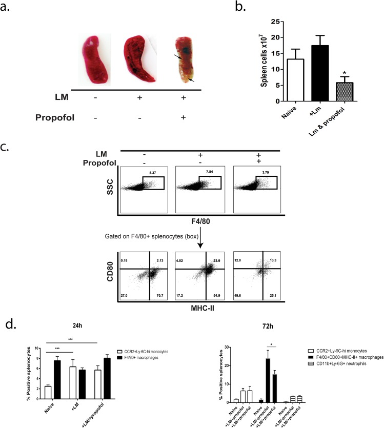
Fig 4. Propofol anesthesia results in loss of splenic architecture and reduced numbers of immune effector cell populations at sites of infection.
(a). Spleens of anesthetized, infected animals were reduced in size and exhibited signs of necrosis at 72 hours post-infection. (b) Total splenocyte counts were lower in propofol-treated animals compared with infected controls. (c). Analysis of cell populations in the spleens of Lm infected animals ± propofol. Swiss Webster mice were left uninfected or infected i.v. with 2 x 10⁴ CFU of Lm in the presence or absence of propofol and spleens were processed for FACS. At 72 hours post-infection, anesthetized animals had proportionally fewer F4/80+CD80+MHC-II+ mononuclear phagocytes than infected controls. (d). At 24 hours post-infection, no significant differences in the proportional numbers of inflammatory monocytes (CCR2+Ly-6Chi) or F4/80+CD80+MHC-II+ mononuclear phagocytes were observed in the spleens of infected animals, regardless of exposure to propofol (left panel). However, at 72 hours post-infection, propofol-treated mice displayed proportionally fewer F4/80+CD80+MHC-II+ mononuclear phagocytes in the spleens compared with infected control animals. No significant changes in the proportion of neutrophils were observed (right panel). *p<0.05, ***p<0.001.

