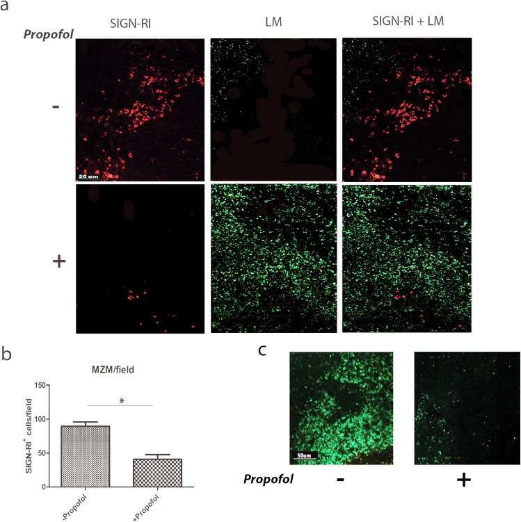
Fig 6. Propofol exposure leads to reduced numbers of marginal zone macrophages and T cell populations in the spleen, and facilitates dissemination of Lm into the white pulp.
Mice were infected with 2 x 10⁴ CFU Lm via intravenous tail vein inoculation in the absence or presence of propofol and sacrificed at 72 hours post-infection. Spleens were harvested, fixed, and antibody-stained for the marginal zone macrophage marker SIGN-RI (red) as well as Lm (green) (a) or for the pan-T cell marker CD3 (green) (c). Images shown are all taken at a 10x magnification and are representative of 3 independent experiments. (b). Quantitation of marginal zone macrophages in the presence or absence of propofol in spleens of Lm-infected animals. Data is an average of 2–4 animals per treatment group, with counts taken from 5 fields per spleen section. Error bars indicate data ± SEM. * p<0.05.

