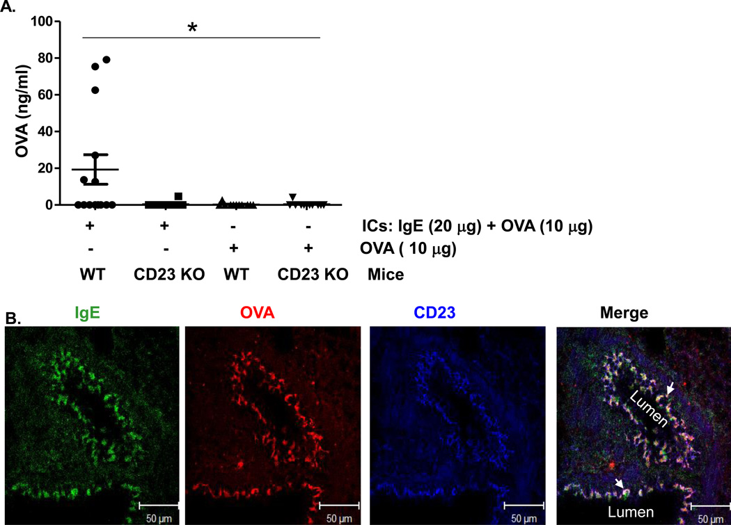
Fig 3. CD23-mediated transcytosis of OVA-IgE immune complexes (ICs) across mouse epithelial monolayers.
(A). Apical to basolateral transcytosis of ICs in naive mice. ICs were formed with 20 µg OVA-specific IgE and 10 µg of OVA at room temperature for 30 min. WT or CD23 KO mice were anaesthetized with avertin and ICs, 10µg OVA, or PBS was i.n. inoculated, respectively. Sera were collected 8 h later and OVA antigen quantitated by ELISA. *P<0.05.
(B). Colocalization of CD23 and ICs in the lungs of naïve mice. OVA-IgE ICs or PBS were i.n. inoculated into anaesthetized mice, which were sacrificed 20 min later and lung tissue was prepared in OCT medium and cryosectioned at 5 µm. Frozen sections were fixed and permeabilized with ice-cold acetone and blocked with 10% NGS. Sections were incubated with rabbit anti-CD23 Ab or mouse anti-chicken OVA Ab followed by staining with Alexa fluor 633-conjugated goat anti-rabbit Ab, Alexa fluor 555-conjugated goat anti-mouse Ab, and FITC conjugated goat anti-mouse IgE Ab. Nuclei were stained with DAPI and sections were imaged using a LSM510 confocal microscope. Samples were visualized under consistent contrast and brightness settings. Arrows indicate the colocalization (white).

